A true educational tool, the creation of educational videos in laparoscopic surgery is subject to certain standards. This includes presenting the clinical case, imaging, the surgical technique, as well as post-operative care and anatomopathological results. Here’s everything you need to know to make an instructive laparoscopic surgery video.

Video-assisted surgery
Laparoscopic surgery is well-adapted for the creation of audiovisual educational materials that enhance the sharing of information through broadcasting channels and within the scientific community
Today, laparoscopic surgery is conducive to creating audiovisual educational materials. These tools facilitate the sharing of information through broadcasting channels and within the scientific community, which fuels debates, technical advancements, helps dissemination of information and communication among surgical teams. In the image of research studies, which produce transparent and reproducible results with a comprehensive and precise report, it is now essential for surgeons to appropriately communicate surgical techniques and their results. Until recently, there were no guidelines for the presentation of surgical videos, resulting in varying quality, reliability, and educational rigor in these documents.
The creation of a laparoscopic surgery video should be a well-defined procedure to make a video both educational and scientifically valid. Recently, an international committee of experts composed of trainers and learners from various specialties drafted and published a consensus on the guidelines for presenting educational videos in laparoscopic surgery (Lap-VEGaS: LAParoscopic surgery Video Educational GuidelineS). The aim of these recommendations was to provide consensus advice on how to present a laparoscopic surgical video for educational and scientific purposes. These recommendations will be briefly presented here.
Laparoscopic Surgery Videos: Best Educational Practices to Follow
A laparoscopic surgery video can become a valuable educational tool by adhering to the following steps:
Author Information and Video Introduction
To begin with, it is important for the video to include author information such as names, institutions, the country, the year of the surgery, and the author’s contact details. The video title should include the names of the procedure performed and the treated pathology, and it should specify if the video was presented at national/international meetings or recorded during a live broadcast. If the video is intended for training, this should be clarified, and the relevance of the case presented, along with specific learning objectives, can be outlined. Lastly, obtaining patient consent is crucial, and a conflict of interest statement should be displayed.
Presentation of the Clinical Case
First and foremost, when creating surgical videos, maintaining the anonymity of the patients is of utmost importance. All radiological images, videos, and reports must be anonymized, and the patient’s name should never be mentioned. Any identifiable parts of the patient’s body, such as eyes and tattoos, must be obscured. If this is impossible, blurring or pixelating the relevant areas is necessary.
The video should include one or more slides or audio comments with a formal presentation of the case, including the patient’s age, gender, American Society of Anesthesiologists (ASA) score, body mass index (BMI), the indication of surgery, comorbidities, and previous surgical history. Preoperative imaging results should be presented, and important anatomical elements, such as potential anatomical or vascular variations, should be mentioned to help the viewer better understand the challenges faced. Blood test results should also be briefly presented if relevant to the case. Finally, as mentioned earlier, caution must be exercised in providing educational context in the video (age, type of pathology, medical history…) because patient re-identification is possible when specific information is combined.
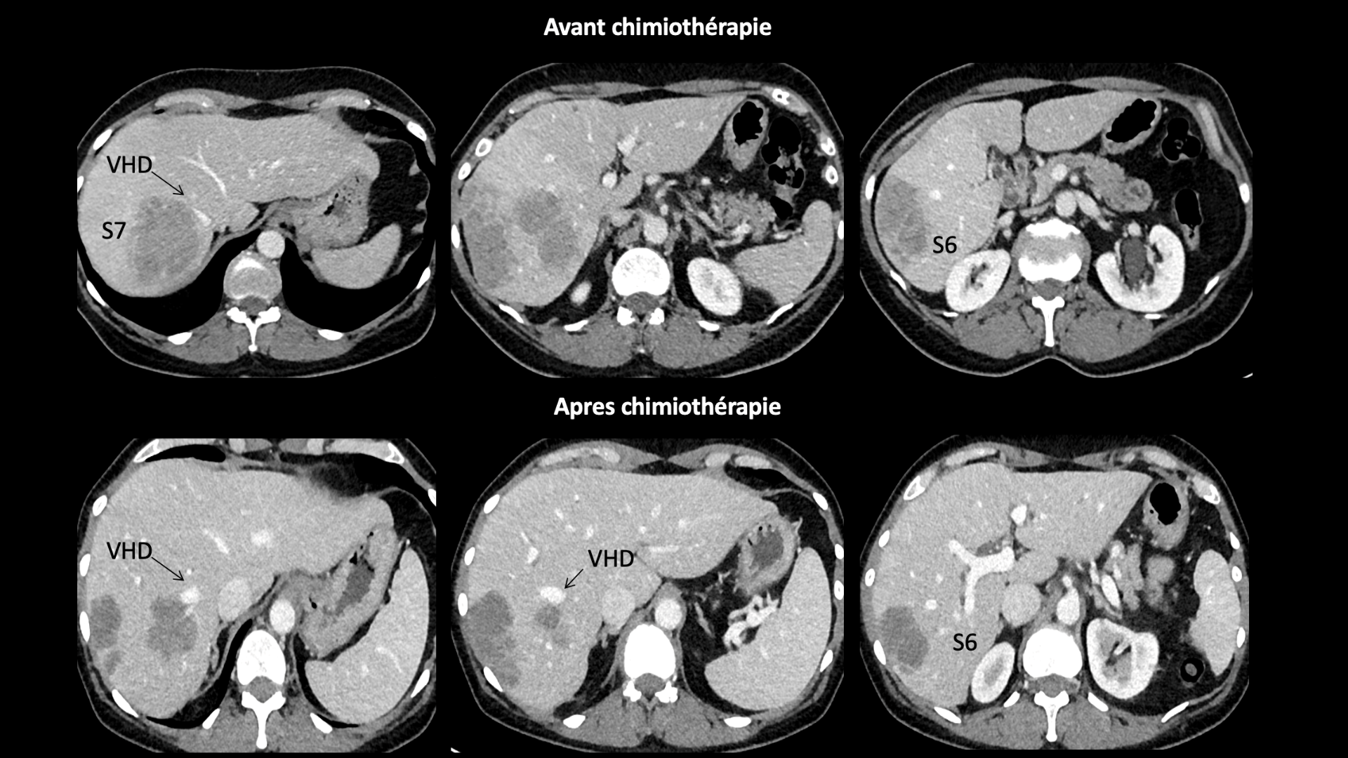
Presentation of the Clinical Case
The video should include one or more slides with a formal presentation of the case and its preoperative imaging. Important anatomical elements should be mentioned to help the viewer better understand the challenges encountered.
Demonstration of the Surgical Procedure
If this part of the video aims to demonstrate the crucial portion of the intra-abdominal surgical procedure, it is often overlooked, yet it is equally important to explain the “outside” environment (extra-peritoneal) since it can vary depending on team preferences.
The initial part of the video should include:
- The patient’s positioning on the operating table, including any changes during the surgery.
- The positioning of the surgical and anesthesia team, including the position of the operating room nurse and additional assistants.
- The detailed placement of trocars. It should be mentioned where additional trocars may be inserted in case of unexpected discoveries or technical difficulties.
- Demonstration of the specimen extraction site.
The intra-peritoneal part of the video should include:
- Details of the special equipment required for the procedure, such as vessel-sealing devices, wound protectors, manipulators, and surgical staplers.
- The surgical procedure should be presented in a standardized step-by-step manner. Intraoperative findings should be demonstrated, with constant reference to anatomy. Graphic elements, like arrows and annotations, can be highly useful in illustrating significant steps during the surgery. These elements are often transparent overlays added to the video image, providing graphical or textual details about aspects of the procedure – anatomy, key actions, and more.
- Additional maneuvers and suggestions for addressing a “progression failure,” such as additional trocar placement or adding an assistant, changes in the patient’s position, or rescue maneuvers in the event of unexpected occurrences, like malfunctioning surgical staplers or equipment failures.
- Description of the criteria for conversion to open surgery and the incision location in case of conversion should also be noted. Similarly, the open portion of the procedure should be mentioned or demonstrated if the video is intended for training.

Demonstration of the Surgical Procedure
The surgical procedure should be presented in a standardized, step-by-step manner with constant reference to anatomy. Graphic elements, such as arrows and annotations, can be of valuable assistance in illustrating important surgical steps.
Results of the procedure
An essential part of an educational video in the field of laparoscopic surgery as well as in any other type of surgery, is to present the results of the procedure. This includes the duration of the operation, blood loss, photos of healed wounds, the length of the hospital stay, and potential postoperative complications.
Another qualitative criterion for assessing laparoscopic surgery, in addition to postoperative outcomes, is the histopathological result of the specimen. This is especially important to detail in cases of malignancy, including the number of lymph nodes removed and staging. Images of the specimen may be desirable.
Adhering to these recommendations can greatly enhance the educational potential of a laparoscopic surgery video, which, after all, is the primary goal one seeks to achieve.
Article written by Stylianos Tzedakis.
Read more
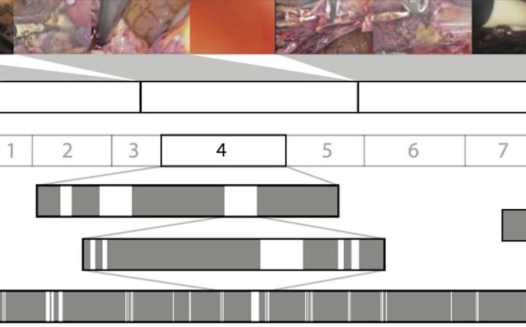
Surgical Video Summarization: Multifarious Uses, Summarization Process and Ad-Hoc Coordination
While surgical videos are valuable support material for activities around surgery, their summarization demands great amounts of time from surgeons, limiting the production of videos. Through fieldwork, we show current practices around surgical videos. First, we...
ResoMeds: a social network sharing videos
Case reports enrich medical knowledge and training, and improve practice [1-4]. However, their publication is often limited in existing journals [5]. We aim to highlight the importance of creating a platform for physician exchange to promote peer learning, case...
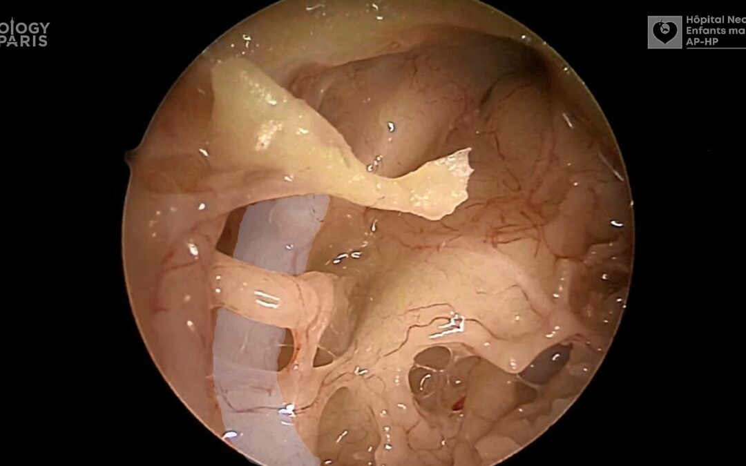
Videos improve knowledge retention of surgical anatomy
In otolaryngology, a new publication shows that an educational video improves anatomy learning and knowledge retention in the long term. This study conducted by the ENT team at Necker-Enfants Malades, APHP (Université Paris Cité) and led by Pr François Simon shows the...
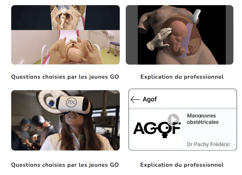
Surgical videos: using the most efficient medium. AGOF’s associative experience
In 2010, the French National Authority for Health (HAS) issued the famous slogan for apprentice surgeons: "Never perform surgery on a patient for the first time" (1). It is sometimes difficult for a young surgeon to accept that he or she has not received sufficient...
